- Home
-
Products
- Magnetic Ionic Liquid
-
Magnetic Beads
- Streptavidin Magnetic Beads
- Aldehyde Magnetic Beads
- Alkynyl Magnetic Beads
- Amino Magnetic Beads
- Avidin Magnetic Bead
- Azide Magnetic Beads
- Carboxyl Magnetic Beads
- DEAE Magnetic Beads
- Epoxy Magnetic Beads
- Heparin Magnetic Beads
- Hydroxyl Magnetic Beads
- NHS Magnetic Beads
- Phenyl Magnetic Beads
- Protein Magnetic Beads
- Silica Magnetic Beads
- Silicon Magnetic Beads
- Thiol Magnetic Beads
- Tosyl Magnetic Beads
- Magnetic Nanodispersion
- Ferrite Magnetic Nanopowder
- High-Purity Magnetic Nanomaterials
- Magnetic Polystyrene Microspheres
- Magnetic Bead Kit
- Magnetic Iron Oxide Nanocrystals
-
Magnetic Nanoparticles
- Amino-Functionalized Magnetic Nanoparticles
- Carboxyl-Functionalized Magnetic Nanoparticles
- Epoxy-Functionalized Magnetic Nanoparticles
- Hydrophobic Magnetic Nanoparticles
- IDA-Functionalized Magnetic Nanoparticles
- Magnetic Fe2O3 Nanoparticles
- Magnetic Fe3O4 Nanoparticles
- PEI Magnetic Nanoparticles
- PVA Magnetic Nanoparticles
- Silanol-Functionalized Magnetic Nanoparticles
- Urea Formaldehyde Magnetic Nanoparticles
- PAMAM Magnetic Nanoparticles
- Protein A Magnetic Nanoparticles
- Protein G Magnetic Nanoparticles
- Streptavidin Magnetic Nanoparticles
- Magnetic Silica Microspheres
-
Services
- Biomagnetic Material
- Magnetic Hydrogel Services
- Rare Earth Magnetic Materials Services
- Magnetic Ionic Liquid Services
- Magnetic Nanomaterials Services
- Magnetic Activated Carbon Service
- Molecular Magnetic Materials Services
- Magnetic Compounds Services
-
Custom Magnetic Beads Services
-
Biofunctionalized Magnetic Bead Services
- Magnetic Bead Protein Conjugation Services
- Magnetic Bead Antibody Conjugation Services
- Magnetic Bead Aptamer Conjugation Services
- Magnetic Bead Carbohydrate Conjugation Services
- Magnetic Bead Nucleic Acid Conjugation Services
- Magnetic Bead Drug Conjugation Services
- Magnetic Bead Enzyme Conjugation Services
- Magnetic Bead Metal Conjugation Services
- Magnetic Bead Multifunctional Composite Conjugation Services
- Magnetic Bean Affinity Tag Conjugation Services
- Magnetic Bead Affinity Ligand Conjugation Services
- Magnetic Bead Antigen Conjugation Services
- Magnetic Bead Fluorescent Material Conjugation Services
- Magnetic Bead Multiplex Conjugation Services
- Magnetic Bead Stimulus-Responsive Modification Services
- Magnetic Bead Enzyme-Responsive Modification Services
- Magnetic Bead Peptide Conjugation Services
- Magnetic Beads Glycolipid Conjugation Services
- Magnetic Beads Glycoprotein Conjugation Services
- Magnetic Bead Fluorescent Dye Conjugation Services
- Fluorescent-Magnetic Dual Functional Beads
- Magnetic Bead Conjugation Service for CRISPR Ribonucleoprotein
- Multiplexed Encoded Magnetic Beads Design Service
- Custom Magnetic Bead Synthesis
-
High-Performance Magnetic Beads Service
- Exosome Magnetic Bead Services
- High Affinity Magnetic Bead Service
- Immobilized Enzyme Magnetic Bead Services
- Low Non-Specific Adsorption Magnetic Bead Service
- Magnetic Bead Microfluidics Services
- Multimodal Magnetic Bead Services
- Protein Crystallization Magnetic Bead Services
- Ultra-Fast Responsive Magnetic Bead Services
- High-Stability Magnetic Bead Services
- Automated Magnetic Bead Processing Service
- Biocompatible Magnetic Bead Design Service
- Cell Sorting Magnetic Bead Design Service
- High Performance Magnetic Bead Characterization Service
- High-Sensitivity Biomarker Magnetic Bead Service
- Immune Enrichment Magnetic Bead Service
- Magnetic Bead GMP Production Service
- Molecular Diagnostics Magnetic Bead Service
- Nucleic Acid Extraction Magnetic Bead Design Service
- Protein Purification Magnetic Bead Design Service
- Protein Purification Magnetic Bead Service
-
Biofunctionalized Magnetic Bead Services
-
Nanomedicine Service
-
New Nanoparticle Design Services
- Complex Nanoparticle Design
- Nanoparticle Scalability Testing Service
- Polymer Nanoparticle Design Services
- Biobased Nanoparticles Design Service
- Gold Nanomaterials Design Service
- Metallic Nanoparticles Design Service
- Magnetite Nanoparticle Design Service
- Semiconductor Nanoparticles Design Service
- Titanium Dioxide Nanoparticles Design Service
- Metal Oxide Nanoparticles Design Service
- Silica Nanomaterials Design Service
- Carbon-Based Nanoparticles Design Service
- Nanomedicine CDMO Services
- Custom Nanoparticle Development and Manufacturing for Diagnostics
- Custom Nanoparticles Synthesis
- Nanoparticle Characterization Service
-
New Nanoparticle Design Services
-
Magnetic Beads Separation & Purification Services
- Antigen Sorting Magnetic Bead Service
- Clinical Sample Testing Magnetic Bead Service
- Environmental detection Magnetic Bead Service
- Food Testing Magnetic Bead Service
- Protein Sorting Magnetic Bead Service
- Serum Testing Magnetic Bead Service
- Virus Particle Enrichment Magnetic Bead Service
- Pollutant Detection Magnetic Bead Service
-
Magnetic Beads QC & Validation Services
- Magnetic Bead Activity Verification Service
- Magnetic Bead Adsorption Capacity Evaluation Service
- Magnetic Bead Binding Capacity Testing Service
- Magnetic Bead Biocompatibility Analysis Service
- Magnetic Bead Conjugation Efficiency Detection Service
- Magnetic Bead Consistency Validation
- Magnetic Bead Imaging Analysis Service
- Magnetic Bead Magnetic Response Speed Testing Service
- Magnetic Beads Particle Size Distribution Analysis Service
- Magnetic Bead Magnetic Detection Service
- Magnetic Bead Magnetic Strength Testing Service
- Magnetic Bead Morphology Analysis Service
- Magnetic Bead Stability Assessment Service
-
Magnetic Beads Application Development Services
- Magnetic Bead ELISA Platform Development Service
- Magnetic Bead Microfluidic Platform Development Service
- Molecular Diagnosis and Nucleic Acid Platform Development Services
- Nucleic Acid Probe Screening Magnetic Beads Service
- Magnetic Bead High-Throughput Drug Screening Platform Development
- Magnetic Bead Assisted Single-Cell Sorting Platform Development
- Magnetic Bead Based Environmental Pollutant Detection Platform Development
- Magnetic Bead Bubble Separation Platform Development
- Magnetic Bead Clinical Biochemical Index Detection Platform Development
- Magnetic Bead Coupled Vaccine Delivery System Platform Development
- Magnetic Bead Disease Marker Screening Platform Development
- Magnetic Bead Enhanced Chemiluminescence (ClLA) Detection Platform Development
- Magnetic Bead Environment Microbial Research Platform Development
- Magnetic Bead Food Microbial Detection Platform Development
- Magnetic Bead Gene Editing Verification Platform Development
- Magnetic Bead Heavy Metal Detection Platform Development
- Magnetic Bead Inflammatory Marker Detection Platform Development
- Magnetic Bead Liquid Biopsy Platform Development
- Magnetic Bead Pesticide Residue Detection Platform Development
- Magnetic Bead Stem Cell Differentiation Research Platform Development
- Magnetic Bead Stem Cell Screening Platform Development
- Magnetic Bead Tumor Single-Cell Sequencing Platform Development
- Magnetic Bead Virus Vector Purification Platform Development
- Magnetic Bead-Based Foodborne Pathogen Detection Platform Development
- Magnetic Bead Infectious Disease Diagnosis Platform Development
- Magnetic Bead-Based Western Blot Assistance Platform Development
- Amino Blocking Magnetic Bead Design Service
-
Advanced Magnetic Materials Services
- Custom Magnetic Carbon Material Services
-
Magnetic Ionic Liquids and Functional Compounds Service
- Magnetic Compounds Development Services
- Magnetic Ionic Liquid Technology Services
- Magnetic Ionic Liquid Bio-application Services
- Magnetic Ionic Liquid Characterization and Analysis Services
- Magnetic Ionic Liquid Customization Services
- Magnetic Ionic Liquid Formulation Optimization
- Magnetic Ionic Liquid Recycling and Regeneration Services
- Magnetic Ionic Liquid Separation Technology Development Services
- Magnetic Ionic Liquids and Catalysis Services
- Magnetic Ionic Liquid Formulation Optimization Services
- Magnetic Ionic Liquid Analytical Testing Services
- Custom Magnetic Nanoparticle Services
-
Rare Earth Magnetic Materials Development and Coating Services
- Custom Rare Earth Magnetic Material Formulation Services
- High-Performance Rare Earth Magnet Research Services
- Next-Generation Rare Earth Magnet Development Services
- Precision Rare Earth Magnetic Material Engineering
- Rare Earth Magnetic Composite Development Services
- Rare Earth Magnetic Material Coating Service
- Rare Earth Magnetic Materials Property Optimization Services
- Rare Earth Magnetic Materials Technology Services
- Specialty Rare Earth Magnetic Materials Design
- Sustainable Rare Earth Magnet Development Solutions
-
Magnetic Hydrogel and Intelligent Response System Services
- 3D Printing Magnetic Hydrogel Customization Services
- Antibacterial Magnetic Hydrogel Services
- Bio-Application Targeted Magnetic Hydrogel Solutions
- Biomedical Magnetic Hydrogels Services
- Biomedical-Grade Targeted Magnetic Hydrogel Platform Development
- Biotargeted Release Magnetic Hydrogel Services
- Custom Biotargeted Release Magnetic Hydrogel Services
- Custom Multifunctional Magnetic Hydrogel Services
- Drug Delivery Magnetic Hydrogel Development Services
- Enzyme-Responsive Magnetic Hydrogel Platform Development
- Gene Therapy Magnetic Hydrogel Services
- Intelligent Magnetic-Responsive Hydrogel Services
- Magnetic Hydrogel Performance Optimization Services
- Magnetic Hydrogel Synthesis Services
- Magnetic Nanocomposite Hydrogel Customization Services
- Magnetic-Responsive Hydrogel Formulation Design Services
- Microfluidic Magnetic Hydrogel Services
- PH Responsive Magnetic Hydrogel Platform Development
- Thermal Responsive Magnetic Hydrogel Platform Development
-
Magnetic Bead Labeling Technology Service
- Antigen Magnetic Beads Custom Labeling Service
- Antibody Purification Magnetic Bead Design Services
- High-Sensitivity Antibody Magnetic Bead Coating Service
- Antimicrobial Peptide (AMP) Screening Magnetic Bead Service
- Immunomagnetic Bead Oriented Labeling Service
- Capture Sequencing Magnetic Bead Design Service
- Magnetic Bead Biological Ligand Affinity Purification Labeling Service
- Chemiluminescence Magnetic Bead Design Service
- Magnetic Bead Probe Design and Validation Service
- Immunoassay Magnetic Bead Design Service
- Magnetic Bead Surface Carboxyl Activation Service
- Magnetic Bead Agar Modification Service
- Magnetic Beads Labeling Process Development Service
- Magnetic Bead Aldehyde Modification Services
- Multi-Omics Sample Pretreatment Magnetic Bead Labeling Service
- Magnetic Bead Amine Modification Service
- Nucleic Acid Probe Magnetic Bead Conjugation Service
- Magnetic Bead Epoxidation Modification Service
- Magnetic Bead Organic Solvent Resistant Modification Service
- Magnetic Bead Silicon Hydroxy Acid Modification Service
- Magnetic Bead-Based Glycan Enrichment Service
- Magnetic Bead-Based Glycoprotein Enrichment Service
- Magnetic Beads for Host Cell Protein (HCP) Removal Service
- Mitochondria Isolation Magnetic Beads Development
- Nanobody Magnetic Bead Design Services
- Phosphopeptide Enrichment Magnetic Bead Service
- RNA-Seq Magnetic Bead Design Service
- RT-PCR Amplification Magnetic Bead Design
- Solid Phase Extraction Magnetic Bead Development Service
- Thermal Contrast Imaging Magnetic Bead Design Service
- Vaccine Antigen Purification Magnetic Beads Design
- Anti-Biotin Magnetic Bead Development
-
Magnetic Bead Detection Service
- Functional Binding Assessment for Magnetic Beads
- Magnetic Bead Biological Stability Detection Service
- Magnetic Bead Chemical Stability Detection Service
- Magnetic Bead Density Quantification Service
- Magnetic Bead Magnetic Response Speed Detection Service
- Magnetic Bead Sterility Testing Service
- Magnetic Bead Surface Topography & Smoothness Detection Service
- Magnetic Magnetization Detection Service
- Magnetic Size & Distribution Detection Service
- Magnetic Bead Formulation Service
- Magnetic-Bead Based OEM Solutions
- Support
- About Us
- Contact Us

- Magnetic Compounds Services
- Molecular Magnetic Materials Services
- Magnetic Activated Carbon Service
- Magnetic Nanomaterials Services
- Magnetic Ionic Liquid Services
- Rare Earth Magnetic Materials Services
- Magnetic Hydrogel Services
- Biomagnetic Material
-
Custom Magnetic Beads Services
-
Biofunctionalized Magnetic Bead Services
- Magnetic Bead Protein Conjugation Services
- Magnetic Bead Antibody Conjugation Services
- Magnetic Bead Aptamer Conjugation Services
- Magnetic Bead Carbohydrate Conjugation Services
- Magnetic Bead Nucleic Acid Conjugation Services
- Magnetic Bead Drug Conjugation Services
- Magnetic Bead Enzyme Conjugation Services
- Magnetic Bead Metal Conjugation Services
- Magnetic Bead Multifunctional Composite Conjugation Services
- Magnetic Bean Affinity Tag Conjugation Services
- Magnetic Bead Affinity Ligand Conjugation Services
- Magnetic Bead Antigen Conjugation Services
- Magnetic Bead Fluorescent Material Conjugation Services
- Magnetic Bead Multiplex Conjugation Services
- Magnetic Bead Stimulus-Responsive Modification Services
- Magnetic Bead Enzyme-Responsive Modification Services
- Magnetic Bead Peptide Conjugation Services
- Magnetic Beads Glycolipid Conjugation Services
- Magnetic Beads Glycoprotein Conjugation Services
- Magnetic Bead Fluorescent Dye Conjugation Services
- Fluorescent-Magnetic Dual Functional Beads
- Magnetic Bead Conjugation Service for CRISPR Ribonucleoprotein
- Multiplexed Encoded Magnetic Beads Design Service
- Custom Magnetic Bead Synthesis
-
High-Performance Magnetic Beads Service
- Exosome Magnetic Bead Services
- High Affinity Magnetic Bead Service
- Immobilized Enzyme Magnetic Bead Services
- Low Non-Specific Adsorption Magnetic Bead Service
- Magnetic Bead Microfluidics Services
- Multimodal Magnetic Bead Services
- Protein Crystallization Magnetic Bead Services
- Ultra-Fast Responsive Magnetic Bead Services
- High-Stability Magnetic Bead Services
- Automated Magnetic Bead Processing Service
- Biocompatible Magnetic Bead Design Service
- Cell Sorting Magnetic Bead Design Service
- High Performance Magnetic Bead Characterization Service
- High-Sensitivity Biomarker Magnetic Bead Service
- Immune Enrichment Magnetic Bead Service
- Magnetic Bead GMP Production Service
- Molecular Diagnostics Magnetic Bead Service
- Nucleic Acid Extraction Magnetic Bead Design Service
- Protein Purification Magnetic Bead Design Service
- Protein Purification Magnetic Bead Service
-
Biofunctionalized Magnetic Bead Services
-
Nanomedicine Service
- Nanomedicine CDMO Services
- Custom Nanoparticle Development and Manufacturing for Diagnostics
- Custom Nanoparticles Synthesis
- Nanoparticle Characterization Service
-
New Nanoparticle Design Services
- Complex Nanoparticle Design
- Nanoparticle Scalability Testing Service
- Polymer Nanoparticle Design Services
- Biobased Nanoparticles Design Service
- Gold Nanomaterials Design Service
- Metallic Nanoparticles Design Service
- Magnetite Nanoparticle Design Service
- Semiconductor Nanoparticles Design Service
- Titanium Dioxide Nanoparticles Design Service
- Metal Oxide Nanoparticles Design Service
- Silica Nanomaterials Design Service
- Carbon-Based Nanoparticles Design Service
-
Magnetic Beads Separation & Purification Services
- Antigen Sorting Magnetic Bead Service
- Clinical Sample Testing Magnetic Bead Service
- Environmental detection Magnetic Bead Service
- Food Testing Magnetic Bead Service
- Protein Sorting Magnetic Bead Service
- Serum Testing Magnetic Bead Service
- Virus Particle Enrichment Magnetic Bead Service
- Pollutant Detection Magnetic Bead Service
-
Magnetic Beads QC & Validation Services
- Magnetic Bead Activity Verification Service
- Magnetic Bead Adsorption Capacity Evaluation Service
- Magnetic Bead Binding Capacity Testing Service
- Magnetic Bead Biocompatibility Analysis Service
- Magnetic Bead Conjugation Efficiency Detection Service
- Magnetic Bead Consistency Validation
- Magnetic Bead Imaging Analysis Service
- Magnetic Bead Magnetic Response Speed Testing Service
- Magnetic Beads Particle Size Distribution Analysis Service
- Magnetic Bead Magnetic Detection Service
- Magnetic Bead Magnetic Strength Testing Service
- Magnetic Bead Morphology Analysis Service
- Magnetic Bead Stability Assessment Service
-
Magnetic Beads Application Development Services
- Magnetic Bead ELISA Platform Development Service
- Magnetic Bead Microfluidic Platform Development Service
- Molecular Diagnosis and Nucleic Acid Platform Development Services
- Nucleic Acid Probe Screening Magnetic Beads Service
-
Magnetic Bead High-Throughput Drug Screening Platform Development
- Magnetic Bead Antibody Drug Screening Platform Development
- Magnetic Bead Biopharmaceutical Drug Screening Platform Development
- Magnetic Bead Cancer Early Screening Platform Development
- Magnetic Bead Cell Therapy Drug Screening Platform Development
- Magnetic Bead Microbial Drug Screening Platform Development
- Magnetic Bead Nucleic Acid Drug Screening Platform Development
- Magnetic Bead Oligosaccharides Drug Screening Platform Development
- Magnetic Bead Peptide Drug Screening Platform Development
- Magnetic Bead Plant Molecular Breeding Platform Development
- Magnetic Bead Protein Drug Screening Platform Development
- Magnetic Bead Radiopharmaceutical Screening Platform Development
- Magnetic Bead Small Molecule Drug Screening Platform Development
- Magnetic Bead Targeted Drug Screening Platform Development
- Magnetic Bead Assisted Single-Cell Sorting Platform Development
- Magnetic Bead Based Environmental Pollutant Detection Platform Development
- Magnetic Bead Bubble Separation Platform Development
- Magnetic Bead Clinical Biochemical Index Detection Platform Development
- Magnetic Bead Coupled Vaccine Delivery System Platform Development
- Magnetic Bead Disease Marker Screening Platform Development
- Magnetic Bead Enhanced Chemiluminescence (ClLA) Detection Platform Development
- Magnetic Bead Environment Microbial Research Platform Development
- Magnetic Bead Food Microbial Detection Platform Development
- Magnetic Bead Gene Editing Verification Platform Development
- Magnetic Bead Heavy Metal Detection Platform Development
- Magnetic Bead Inflammatory Marker Detection Platform Development
- Magnetic Bead Liquid Biopsy Platform Development
- Magnetic Bead Pesticide Residue Detection Platform Development
- Magnetic Bead Stem Cell Differentiation Research Platform Development
- Magnetic Bead Stem Cell Screening Platform Development
- Magnetic Bead Tumor Single-Cell Sequencing Platform Development
- Magnetic Bead Virus Vector Purification Platform Development
- Magnetic Bead-Based Foodborne Pathogen Detection Platform Development
- Magnetic Bead Infectious Disease Diagnosis Platform Development
- Magnetic Bead-Based Western Blot Assistance Platform Development
- Amino Blocking Magnetic Bead Design Service
-
Advanced Magnetic Materials Services
- Custom Magnetic Carbon Material Services
-
Magnetic Ionic Liquids and Functional Compounds Service
- Magnetic Compounds Development Services
- Magnetic Ionic Liquid Technology Services
- Magnetic Ionic Liquid Bio-application Services
- Magnetic Ionic Liquid Characterization and Analysis Services
- Magnetic Ionic Liquid Customization Services
- Magnetic Ionic Liquid Formulation Optimization
- Magnetic Ionic Liquid Recycling and Regeneration Services
- Magnetic Ionic Liquid Separation Technology Development Services
- Magnetic Ionic Liquids and Catalysis Services
- Magnetic Ionic Liquid Formulation Optimization Services
- Magnetic Ionic Liquid Analytical Testing Services
- Custom Magnetic Nanoparticle Services
-
Rare Earth Magnetic Materials Development and Coating Services
- Custom Rare Earth Magnetic Material Formulation Services
- High-Performance Rare Earth Magnet Research Services
- Next-Generation Rare Earth Magnet Development Services
- Precision Rare Earth Magnetic Material Engineering
- Rare Earth Magnetic Composite Development Services
- Rare Earth Magnetic Material Coating Service
- Rare Earth Magnetic Materials Property Optimization Services
- Rare Earth Magnetic Materials Technology Services
- Specialty Rare Earth Magnetic Materials Design
- Sustainable Rare Earth Magnet Development Solutions
-
Magnetic Hydrogel and Intelligent Response System Services
- 3D Printing Magnetic Hydrogel Customization Services
- Antibacterial Magnetic Hydrogel Services
- Bio-Application Targeted Magnetic Hydrogel Solutions
- Biomedical Magnetic Hydrogels Services
- Biomedical-Grade Targeted Magnetic Hydrogel Platform Development
- Biotargeted Release Magnetic Hydrogel Services
- Custom Biotargeted Release Magnetic Hydrogel Services
- Custom Multifunctional Magnetic Hydrogel Services
- Drug Delivery Magnetic Hydrogel Development Services
- Enzyme-Responsive Magnetic Hydrogel Platform Development
- Gene Therapy Magnetic Hydrogel Services
- Intelligent Magnetic-Responsive Hydrogel Services
- Magnetic Hydrogel Performance Optimization Services
- Magnetic Hydrogel Synthesis Services
- Magnetic Nanocomposite Hydrogel Customization Services
- Magnetic-Responsive Hydrogel Formulation Design Services
- Microfluidic Magnetic Hydrogel Services
- PH Responsive Magnetic Hydrogel Platform Development
- Thermal Responsive Magnetic Hydrogel Platform Development
-
Magnetic Bead Labeling Technology Service
- Antigen Magnetic Beads Custom Labeling Service
- High-Sensitivity Antibody Magnetic Bead Coating Service
- Immunomagnetic Bead Oriented Labeling Service
- Magnetic Bead Biological Ligand Affinity Purification Labeling Service
- Magnetic Bead Probe Design and Validation Service
- Magnetic Bead Surface Carboxyl Activation Service
- Magnetic Beads Labeling Process Development Service
- Multi-Omics Sample Pretreatment Magnetic Bead Labeling Service
- Nucleic Acid Probe Magnetic Bead Conjugation Service
- Antibody Purification Magnetic Bead Design Services
- Antimicrobial Peptide (AMP) Screening Magnetic Bead Service
- Capture Sequencing Magnetic Bead Design Service
- Chemiluminescence Magnetic Bead Design Service
- Immunoassay Magnetic Bead Design Service
- Magnetic Bead Agar Modification Service
- Magnetic Bead Aldehyde Modification Services
- Magnetic Bead Amine Modification Service
- Magnetic Bead Epoxidation Modification Service
- Magnetic Bead Organic Solvent Resistant Modification Service
- Magnetic Bead Silicon Hydroxy Acid Modification Service
- Magnetic Bead-Based Glycan Enrichment Service
- Magnetic Bead-Based Glycoprotein Enrichment Service
- Magnetic Beads for Host Cell Protein (HCP) Removal Service
- Mitochondria Isolation Magnetic Beads Development
- Nanobody Magnetic Bead Design Services
- Phosphopeptide Enrichment Magnetic Bead Service
- RNA-Seq Magnetic Bead Design Service
- RT-PCR Amplification Magnetic Bead Design
- Solid Phase Extraction Magnetic Bead Development Service
- Thermal Contrast Imaging Magnetic Bead Design Service
- Vaccine Antigen Purification Magnetic Beads Design
- Anti-Biotin Magnetic Bead Development
-
Magnetic Bead Detection Service
- Functional Binding Assessment for Magnetic Beads
- Magnetic Bead Biological Stability Detection Service
- Magnetic Bead Chemical Stability Detection Service
- Magnetic Bead Density Quantification Service
- Magnetic Bead Magnetic Response Speed Detection Service
- Magnetic Bead Sterility Testing Service
- Magnetic Bead Surface Topography & Smoothness Detection Service
- Magnetic Magnetization Detection Service
- Magnetic Size & Distribution Detection Service
- Magnetic Bead Formulation Service
- Magnetic-Bead Based OEM Solutions
Magnetic Bead Imaging Analysis Service
The precise characterization of magnetic beads is fundamental to their successful application across biotechnology, medical diagnostics, and pharmaceutical development. While techniques like dynamic light scattering provide valuable average size data, they often lack the visual evidence required to confirm morphological integrity, detect subtle aggregates, or visualize surface features. Imaging analysis delivers this critical visual dimension, transforming numerical data into actionable insights. The ability to directly observe magnetic beads—their size distribution, shape uniformity, surface texture, and dispersion state—is indispensable for troubleshooting production processes, validating quality control, and advancing research and development.
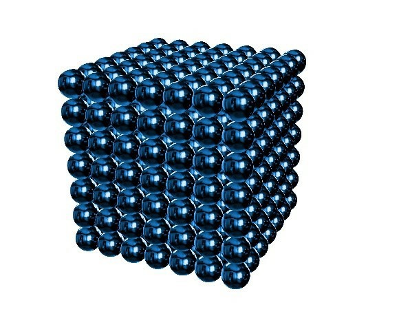 Figure 1. Magnetic Bead.
Overview
CD Bioparticles introduces its comprehensive magnetic bead imaging analysis service, designed to provide clients with high-resolution visual characterization of their magnetic bead formulations. Our service leverages state-of-the-art microscopy technologies to uncover details that other methods cannot detect. We provide more than just images, we deliver detailed analytical reports with quantitative data, offering a clear window into the nano- and micro-scale world of your particles. This service is essential for anyone who needs to confirm the physical structure of their beads, investigate batch inconsistencies, or provide visual evidence for regulatory submissions.
Our Services
CD Bioparticles offers an end-to-end imaging service, from sample preparation to expert image analysis and reporting. Our process is tailored to deliver the specific insights you need.
Consultation and Method Selection
We begin by discussing your objectives. What are your primary concerns? Are you investigating aggregation, verifying size after a process change, or looking at internal structure? Based on this, we recommend the most appropriate imaging technique (SEM, TEM, or a combination) and develop a tailored analysis plan.
Expert Sample Preparation
Sample preparation is critical for obtaining accurate and artifact-free images. Our experienced technicians handle your samples with care, employing optimized protocols:
High-Resolution Imaging
We perform imaging using our advanced microscopy systems, which are maintained to the highest standards. We capture multiple images from different areas of the sample to ensure a representative analysis. We can image beads in a dry state or, using specialized holders, in a liquid suspension for certain analyses.
Comprehensive Image Analysis and Quantification
We go beyond simply providing images. Our service includes detailed quantitative analysis:
Applications
Our Magnetic Bead Imaging Analysis Service is vital for a wide range of applications:
Our Process
Our approach is highly collaborative, ensuring the final product meets your exact specifications:
Consulting and
Design
Synthesis and
functionalization
Characterization and
validation
Quality assurance and
delivery
Our Advantages

Advanced Technology
Our MBI system is built on proprietary technology, including a high-field permanent magnet (1.5 T) for strong magnetic signal generation and a graphene-based magnetic sensor array for ultra-sensitive detection.

Fully Customized Solutions
We reject a "one-size-fits-all" approach. Whether you are a academic researcher studying cell migration in vitro or a pharmaceutical company conducting GLP-compliant in vivo studies, we tailor our service to your specific needs.

Expert Scientific Team
Our team consists of 20+ scientists with backgrounds in biomedical engineering, molecular biology, and imaging technology. This expertise allows us to anticipate challenges and provide proactive solutions, ensuring experiments stay on track.
In the world of magnetic beads, seeing is not just believing—it is understanding. Direct visualization is a powerful tool that complements other analytical techniques, providing unequivocal evidence of bead characteristics that are crucial for performance and reliability. CD Bioparticles is your trusted partner for unlocking these visual insights. Our magnetic bead imaging analysis service provides the clarity and data needed to optimize your products, solve complex challenges, and demonstrate quality with confidence. By partnering with us, you gain access to the expertise and technology necessary to see your magnetic beads in a new light.